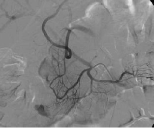

жСЩй20ФъЧАЮвУЧеяЖЯЯћЛЏЕРГібЊвРРЕбЊЙмдьгАЃЈangiographyЃЉЃЌФкПњОЕЃЈendoscopyЃЉКЭКЫЫиЩЈУшЃЈscintigraphyЃЉЁЃЕЋШчЙћУЛгаетаЉвРРЕзЈУХШЫдБВйзїЕФМьВщЪБЃЌдѕУДАьЃП CTAбЊЙмдьгАеяЖЯЯћЛЏЕРГібЊОПОЙЪЧвЛжжЯђЭљЛђПЪЭћЃЌЛЙЪЧЯћЛЏЕРеяЖЯВпТджаЕФвЊЧѓЃЌЩѕжСЧПжЦЁЃ гаЙибЊЙмдьгАЕФРњЪЗЃЈangiography historyЃЉ
CTA ЖдгкЯћЛЏЕРГібЊЕФеяЖЯгжЗжЮЊ CTAПЩвдгУгкШЮКЮаЮЪНЕФЯћЛЏЕРГібЊ
ЯрЙиЮФЯз
Yoon et al. Acute massive GI bleeding: detection and localization with arterial phase MDCT. Radiology, 2006: 239, 160-7
Scheffel et al. Acute GI bleeding: detection of source and etiology with MDCT. Eur Radiol. 2007: 17,1555-65
Ko et al. Localialization of bleeding using MDCT in patietns with signs of acute GI hemorrhage. Rofo 2005: 177, 1649-54
CTбЊЙмдьгАдкеяЖЯМБадЯћЛЏЕРГібЊгагУадЃЈвѕадЕФгагУадЃЌбєадЕФгагУадЃЉШчЯТБэ CTAеяЖЯМБадЯћЛЏЕРГібЊmetaЗжЮіЃЈ2008ЃЉгаЙиЮФЯзМЧдиМБадЯћЛЏЕРCTAЕФНсЙћЃЈP. Goffette, CIRSE 2009ЃЉ
from Jaeckle T, Eur Radiol, 2008, 18:1406 СэЭтвЛЦЊЮФеТПЩПД Diagnostic accuracy of CT angiography in acute gastrointestinal bleedingby Angela Chua and Ridley (Sidney) in J Medical Imaging and Radiation Oncology 2008 CTAеяЖЯМБадЯћЛЏЕРГібЊmetaЗжЮіЃЈ2011ЃЉfrom Wu et al. World Journal of Gastroenterology 2011 copycat 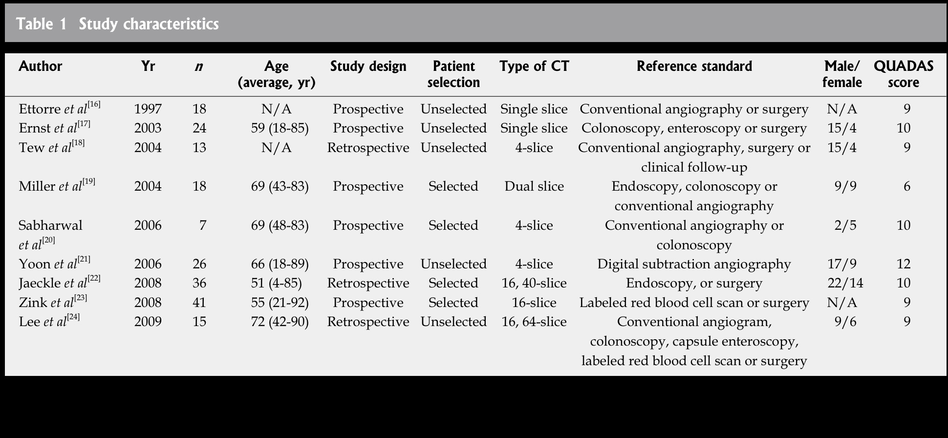
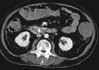
етвЛНЈвщЪЕМЪЩЯИФБфМБадЯћЛЏЕРГібЊЕФСйДВеяЖЯТЗОЖЁЃ 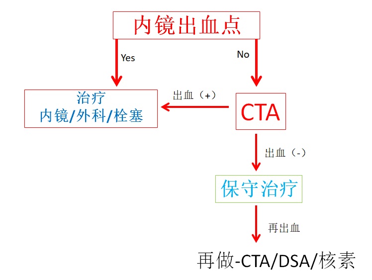 Outcome of negative CTA in acute GIH
°28/46 lower GIH (60%)
CTA ЮЊвѕадЕФВЁР§ЃЌБЃЪижЮСЦЕФ%ИпЁЃ High % of conservative treatment after neg CTA
CTAвѕадЕФМБадЯТЯћЛЏЕРГібЊЃЌНщШыЕФФаХЎУЧЃЌЫЦКѕПЩвдРСДВЃЌЛђДђИпЖћЗђЛђЙКЮя
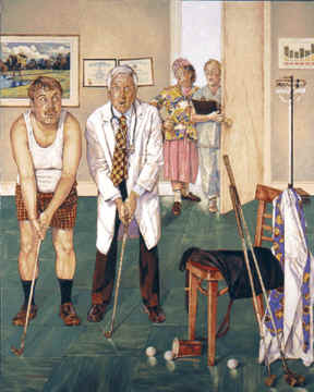
IR man or woman,ЃЌit seems you can stay in bed, or go on golfing, or shopping
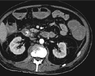
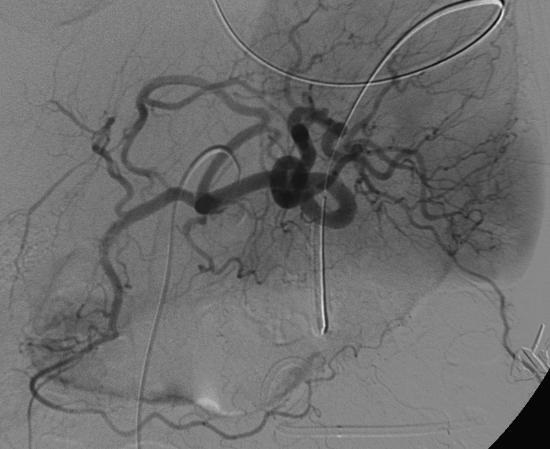
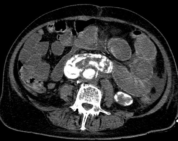
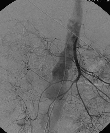
МБадЮИГІЕРГібЊCTA бєадЕФНсЙћЃЈOutcome of positive CTA in acute GIHЃЉЃЌЮвУЧФмЙЛИљОнМБадЯћЛЏЕРГібЊCTAбєадЕФНсЙћОіЖЈЪЧЗёЛМепашвЊЭтПЦЃЌЛђдйДЮФкОЕжЮСЦЃЌЛђбЊЙмдьгАЃЌЛђепШдШЛЕШД§ 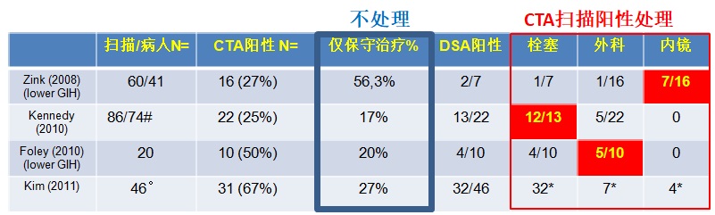
* not specified whether in post/neg CTA
# 59/74 lower GIH (80%)
°28/46 lower GIH (60%)
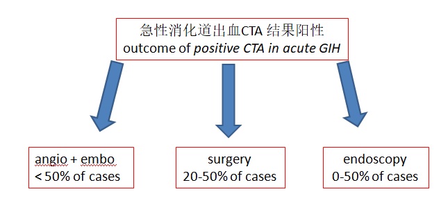 ЩѕжСдкCTAбєадЕФЧщПіЯТЃЌвВВЂУЛгавЛИізМШЗЕФжИФЯПЩЬсЙЉЃЌЕЋНщШыФаХЎУЧгІИУПЊЪМЪБПЬзМБИРВЃЁ
there is no clear algorithm, even in positive CTA cases, but be ready to intervene, IR man or woman
 КЫЫиЩЈУшгыCTAЃЌЮвУЧгІИУСЊКЯЫќУЧТ№ЃП
Zink et al “Disagreement between Tc-99m RBC and CTA for acute lowerGIH” 2008, AJR
* angio + in 2/10ЃЛembo in 1; surgery in 2; conservative 8/11
ОЁЙмCTAКЭКЫЫиЩЈУшБШНЯВЛвЛжТЃЌЕЋШчЙћНіНіКЫЫиЩЈУшбєадЃЌКмЖрВЁШЫЫЦКѕБЃЪижЮСЦИќКЯЪЪ....ЃЌЫљвд
дкШеГЃЙЄзїжаЃЌМБадЯћЛЏЕРГібЊЃЌCTAЪЧЗёгІИУГЩЮЊГЃЙцЃПвЛжжЙлЕуШЯЮЊЃЌзіCTAЮвУЧВЛЛсЪЧдкРЫЗбЪБМфТ№ЃП ЫЦКѕВЂВЛШчДЫЃЌКмЖрCTAЪЧвѕадЕФЃЌгаЪБМфОіЖЈЫЈШћ/ЭтПЦ/ФкОЕЁЃЕБCTAЪБвѕадЃЌВЂВЛашвЊЛ§МЋЕиНјаагаДДЕФжЮСЦЃЌПЩвдЕШзХЧЦЃЈwait and see...ЃЉЁЃЮвУЧжЛашЬзЕЅаадьгАМСЕМжТЕФЩіЫ№ЩЫЃЌКЭЕчРыЗјЩфЕФгАЯьЁЃОЁЙмЗЂЩњТЪЕЭЃЌЖјЧвгАЯьЪЧЧсЮЂЕФЁЃ МБадЯћЛЏЕРГібЊЕФЗНЗЈбЇ Kim et al, J Comput Assist Tomogr. 2011
ЮвУЧашвЊШЋВПЖМдіЧПТ№ЃПDo we need all enhanced series? 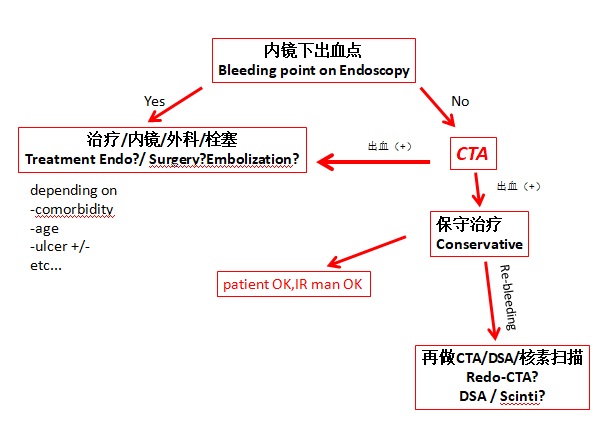 P Goffette CIRSE 2009 Т§адКЭВЛУїдвђЃЈвўФфадЃЉЯћЛЏЕРГібЊКЭCTAЃЈdo you see CTA?ЃЉ
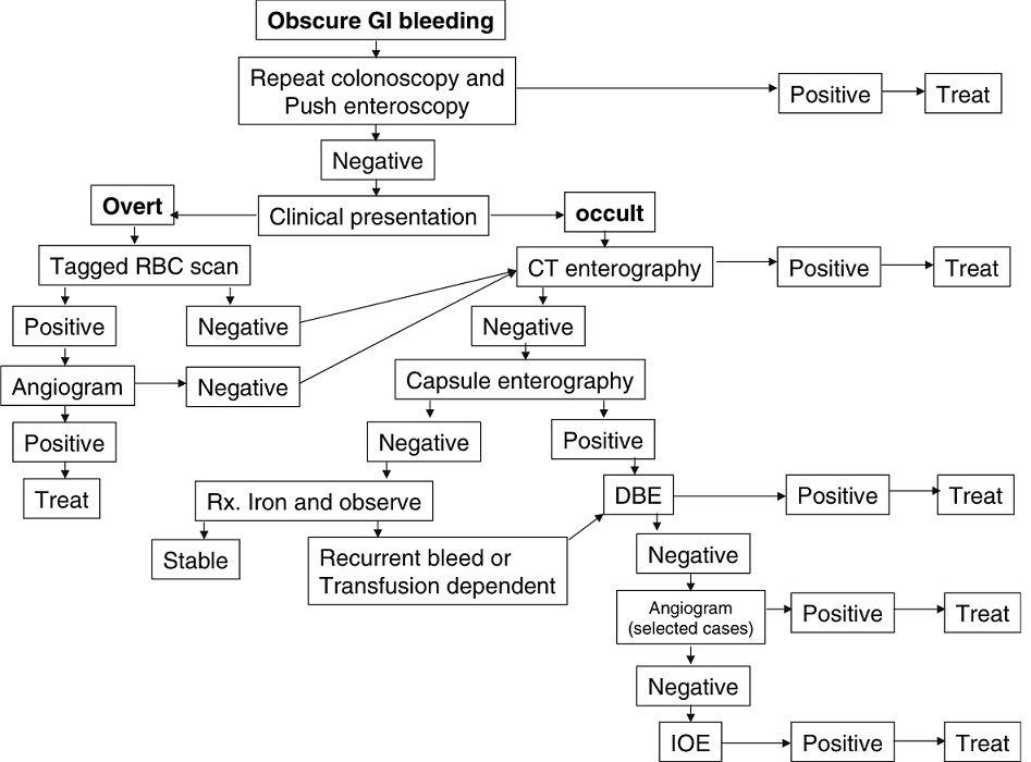
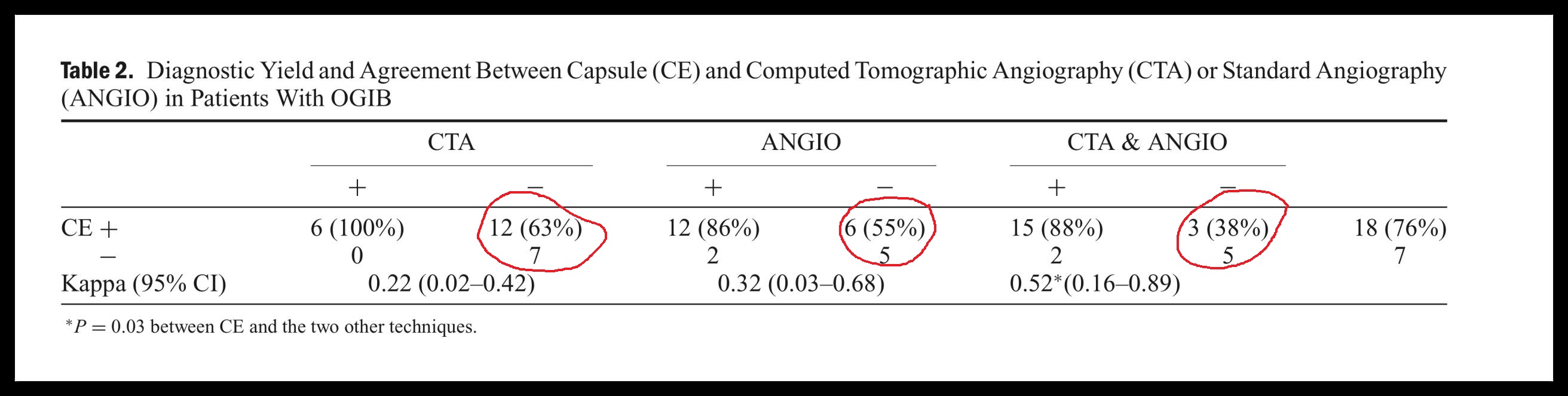
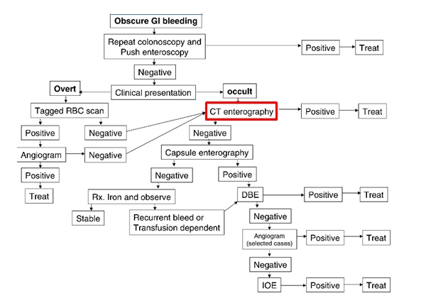
дкТ§адЛђвўФфадЯћЛЏЕРГібЊЕФеяЖЯТЗОЖжаЃЌЩѕжСПДВЛГіCTAЕФзїгУ CTA and chronic/occult/obscure GIH
CTA and chronic/occult GIHЃЈHuprich et al, Radiology, Sept 2011ЃЉ
CTEЩЈУшММЪѕКЭCTAММЪѕЯрЫЦЃЌ64-128ХХЃЌШ§ЦкCTAЃЛ64 x 0.6mm overlapping projectionsКЭMPRжиНЈЁЃ
ВЛУїдвђЕФЯћЛЏЕРГібЊЮЊЪВУДВЛФмСЊКЯCTAЕФЖЏТідьгАЃПObscure GIH: why not combining arteriography with CTA?
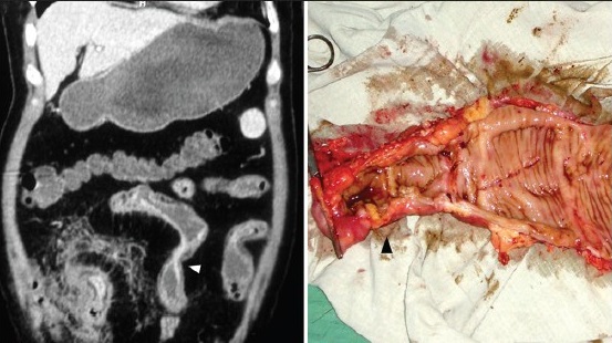
CTA-Mesentericography for obscure GIH
retrospective: 2002-2006 N=6
prospective: 2006-2009 N=7
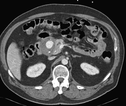
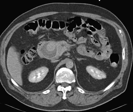
1. жїЖЏТі-ЪЎЖўжИГІ№ќ YES“we feel that MDCTM cannot be recommended in general...” ЮИГІЕРГібЊЪЧЗёУПИіШЫЖМашвЊCTAЃП GI-hemorrhage; Should all patients be imaged with CTA? CTA technique of first choice, sens>90%, spec.>80%
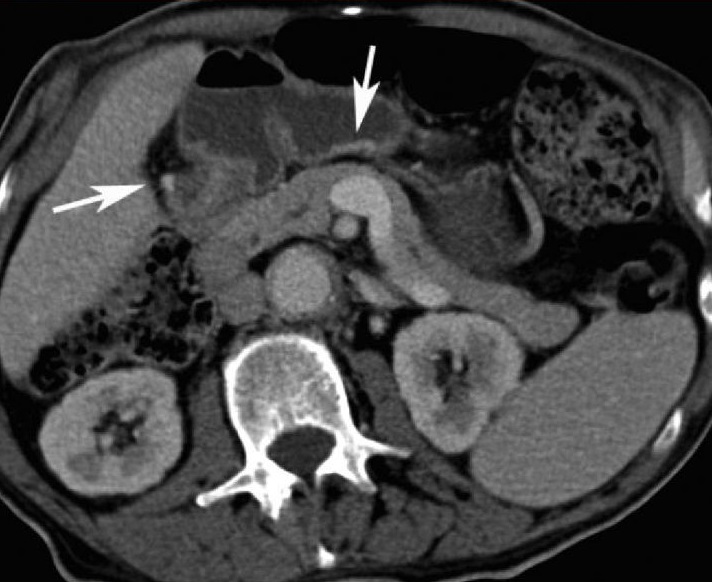
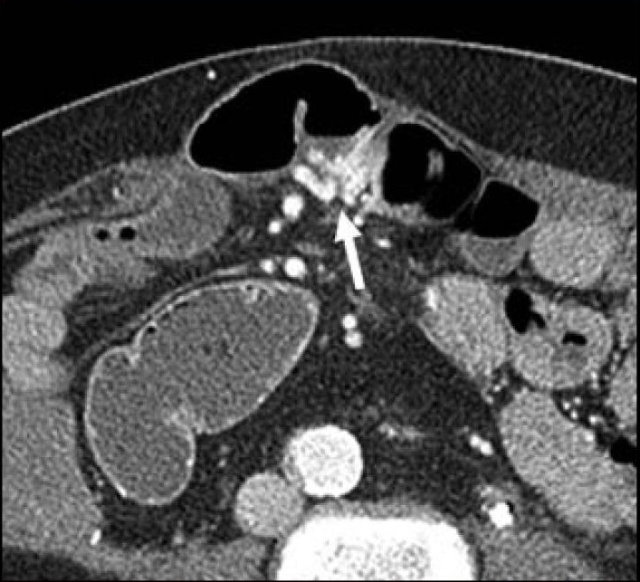
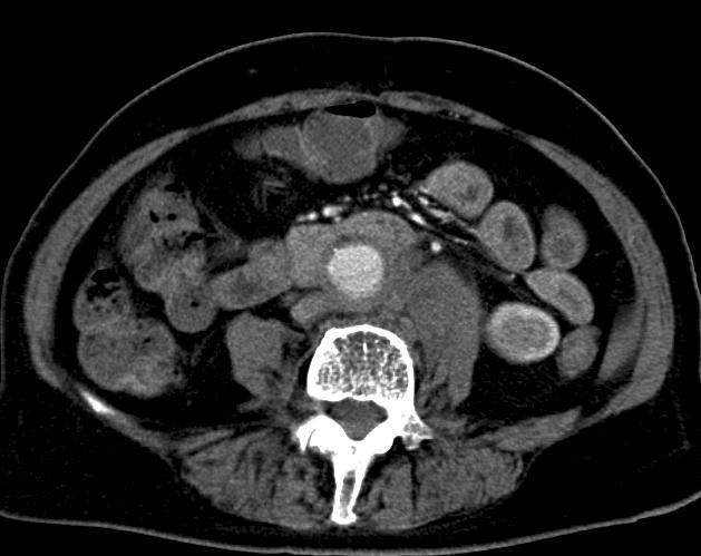
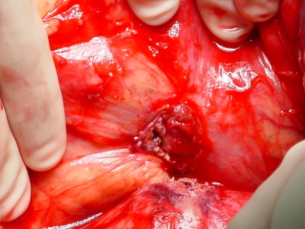
Signs: contrast extravastion in duodenum, peri-graft fluid,
peri-graft soft tissue mass, peri-graft air, focal bowel wall
thickening, absence fatplane vessel wall - bowel wall
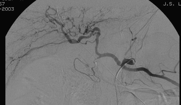
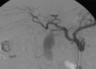
2. ЕЈЕРГібЊ/вШЯйГібЊ Hemobilia / hemosuccus pancreaticus
CTA technique of first choice
Endoscopy limited value for diagnosis and treatment
Embolization treatment of choice
CTA for treatment planning
Hyare et al. MDCT CT angiography compared with DSA in diagnosing major arterial hemorrhage in inflammatory pancreatic disease. Eur J Radiol. 2006: 59, 295-300
3. ЦфЫќдвђ MaybeЃЈПЩФмашвЊНјааCTAЃЉ
Endoscopy more often diagnostic in upper than lower GI tract*
Risk factors for CIN present?
Radiation risk (young patient)?
*Most common causes: upper GI bleed = ulcer bleed ЃЛ lower GI bleed = diverticular bleed
ЮФЯзжаCTAеяЖЯЯћЛЏЕРГібЊЕФНсЙћ 1. ЖЏЮяЪЕбщ
Animal study GI hemorrhage in pigs
CT detection of hemorrhage > 0.3 ml./min.
(Angio detection GI hemorrhage > 0.5 ml./min.)
Kuhle et al. Detection of active colonic hemorrhage with use of helical CT: findings in a swine model.Radiology 2003; 228: 743-52.
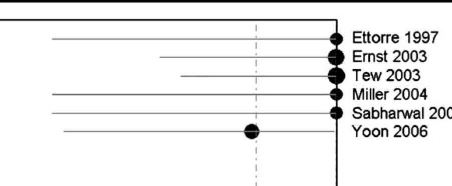
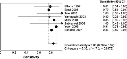
2. Literature results CTA -review
Pooled sens. 86% (95% CI 78–92%)
Pooled spec. 95% (95% CI 76–100%)
Chua et al. Diagnostic accuracy of CTA in acute GI bleeding. J Med Imaging Radiat Oncol. . 2008; 52: 333-8
4. ЛиЙЫадбаОП3. DSAзїЮЊВЮПМБъзМЃЌ50Ч¨ШЫ
Pooled sens. 88%
Retrospective study, 86 CTA’s in 76 pts.with acute GIH
Sens. 79%, spec. 95%, accuracy 91%
PPVЃЈбєаддЄВтжЕЃЉ 86%, NPVЃЈвѕаддЄВтжЕЃЉ 92%
Kennedy et al Active GIH with CTA: a 41/2-year retrospective review. . JVIR 2010; 21: 848-55
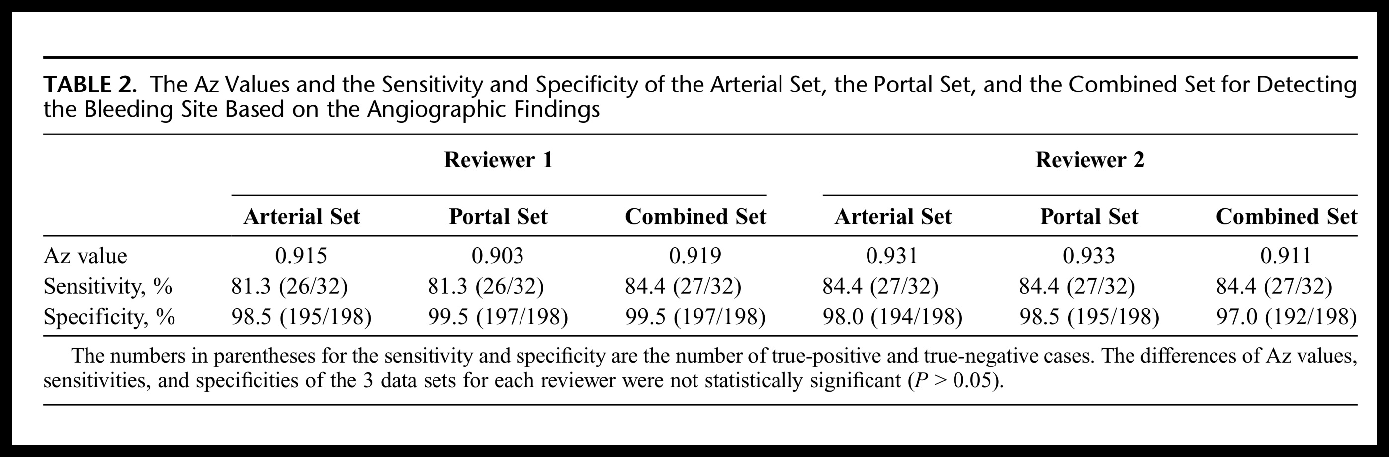
Retrospective study in 26 pts. with confirmed acute GIH
Sens. 92% (24/26)
Jaeckle et al Detection and localization of acute upper and lower GI bleeding with arteria lphase MDCT. . Eur Radiol. 2008; 18: 1406-13
| ||||||||||||||||||||||||||||||||||||||||||||||||||||||||||||||||||||||||||||||||||||||||||||||||||||||||||||||||||||||||||||||||||||||||||||||||||||||||||||||||||||||||||||||||||||||||